Ectopic pituitary adenoma

Ectopic pituitary adenoma
Image Source-Google | Image by- | link.springer 


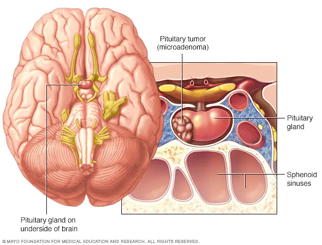
An ectopic (happening in an abnormal vicinity) pituitary adenoma is a rare kind of tumor which takes place outdoor of the sella turcica, most often inside the sphenoid sinus, suprasellar location, nasopharynx and the cavernous sinuses.

Pituitary incidentalomas

Pituitary incidentalomas
Image Source-Google | Image by- | hcplive


Pituitary incidentalomas are pituitary tumors which can be characterized as an incidental finding. They are often found by way of computed tomography (CT) or magnetic resonance imaging (MRI), carried out within the assessment of unrelated scientific conditions including suspected head trauma, in most cancers staging or in the assessment of nonspecific signs which includes dizziness and headache. It isn't unusual for them to be located at autopsy. In a meta-evaluation, adenomas were discovered in an average of 16.7% in postmortem studies, with most being microadenomas (<10mm); macrodenomas accounted for only 0.16% to 0.2% of the decedents. While non-secreting, noninvasive pituitary microadenomas are typically taken into consideration to be actually as well as clinically benign, there are to date scant studies of low best to help this announcement.

It has been advocated in the current Clinical Practice Guidelines (2011) with the aid of the Endocrine Society - a expert, international medical business enterprise inside the area of endocrinology and metabolism - that each one sufferers with pituitary incidentalomas go through a entire medical history and physical examination, laboratory critiques to display screen for hormone hypersecretion and for hypopituitarism. If the lesion is in near proximity to the optic nerves or optic chiasm, a visual view examination ought to be achieved. For those with incidentalomas which do not require surgical elimination, comply with up medical tests and neuroimaging ought to be done as nicely observe-up sight view examinations for incidentalomas that abut or compress the optic nerve and chiasm and comply with-up endocrine checking out for macroincidentalomas.

Classification of Pituitary adenoma

Classification of Pituitary adenoma
Image Source-Google | Image by- | radiopaedia

  • Unlike tumors of the posterior Pituitary, Pituitary adenomas are categorized as endocrine tumors (not mind tumors). Pituitary adenomas are categorized based upon anatomical, histological and functional standards.
  • Anatomically pituitary tumors are categorized by their size based on radiological findings; both microadenomas (much less than <10 mm) or macroadenomas (equal or extra than ≥10 mm).

Classification based on radioanatomical findings locations adenomas into 1 of 4 grades (I–IV):

  • Stage I: microadenomas (<1 cm) with out sella growth.
  • Stage II: macroadenomas (≥1 cm) and may enlarge above the sella.
  • Stage III: macroadenomas with growth and invasion of the ground or suprasellar extension.
  • Stage IV: is destruction of the sella.
  • Histological classification utilizes an immunohistological characterization of the tumors in phrases of their hormone manufacturing. Historically they had been classed as either basophilic, acidophilic, or chromophobic on the idea of whether or not or not they took up the tinctorial stains hematoxylin and eosin. This type has fallen into disuse, in want of a type based totally on what sort of hormone is secreted by way of the tumor. Approximately 20-25% of adenomas do not secrete any effectively identifiable lively hormones ('non-functioning tumors') yet they're still from time to time called 'chromophobic'.
  • Functional type is primarily based upon the tumors endocrine interest as decided with the aid of serum hormone tiers and pituitary tissue cell hormone secretion detected via immunohistochemical staining. The "Percentage of hormone manufacturing cases" values are the fractions of adenomas producing every associated hormone of each tumor type compared to all instances of pituitary tumors, and does not immediately correlate to the odds of every tumor kind due to smaller or extra incidences of absence of secretion of the anticipated hormone. Thus, non secretive adenomas may be either null mobile adenomas or a more specific adenoma that, however, stays non-secretive.
  • Any form of pituitary adenocarcinoma indexed within the desk below may additionally reason compressive signs and symptoms due to nearby growth similarly to the systemic outcomes of secreted hormones indexed within the pathology column.
  • Null cell adenomas by means of definition do now not secrete hormones, however they typically purpose compressive effects at the pituitary stalk (stalk effect). This leads to reduced stages of dopamine from the hypothalamus attaining the anterior pituitary gland. Dopamine exerts an inhibitory impact on prolactin secretion. With the absence of this inhibitory effect, prolactin ranges boom and are frequently expanded in null cell adenomas. This ends in signs of hypogonadism.

Mechanism of Pituitary adenoma

Mechanism of Pituitary adenoma
Image Source-Google | Image by- | researchgate


The pituitary gland or hypophysis is regularly referred to as the "grasp gland" of the human frame. Part of the hypothalamic-pituitary axis, it controls most of the frame's endocrine capabilities thru the secretion of numerous hormones into the circulatory system. The pituitary gland is positioned under the mind in a melancholy (fossa) of the sphenoid bone called the sella turcica. Although anatomically and functionally related to the mind, the pituitary gland sits outdoor the blood–mind barrier. It is separated from the subarachnoid space by the diaphragma sella, consequently the arachnoid mater and as a consequence cerebral spinal fluid can not enter the sella turcica.

The pituitary gland is split into two lobes, the anterior lobe (which debts for two thirds of the extent of the gland), and the posterior lobe (1/3 of the quantity) separated by way of the pars intermedia.

The posterior lobe (the neural lobe or neurohypophysis) of the pituitary gland isn't, in spite of its name, a true gland. The posterior lobe includes axons of neurons that expand from the hypothalamus to which it's miles connected thru the pituitary stalk. The hormones vasopressin and oxytocin, produced via the neurons of the supraoptic and paraventricular nuclei of the hypothalamus, are stored in the posterior lobe and launched from axon endings (dendrites) inside the lobe.

The pituitary gland's anterior lobe (adenohypophysis) is a real gland which produces and secretes six distinct hormones: thyroid-stimulating hormone (TSH), adrenocorticotropic hormone (ACTH), follicle-stimulating hormone (FSH), luteinizing hormone (LH), growth hormone (GH), and prolactin (PRL).

Genetics of FIPA

Genetics of FIPA
Image Source-Google | Image by- | ncbi.nlm.nih

FIPA has  regarded genetic causes, mutations within the AH receptor-interacting protein (AIP) gene and duplications in chromosome Xq26.Three that encompass the GPR101 gene that also causes X-connected acrogigantism (X-LAG) syndrome. About 15-20% of FIPA households deliver a germline AIP gene mutation or deletion, and the disease occurs as autosomal dominant with incomplete penetrance, meaning that approximately 20% of AIP mutation companies will broaden a pituitary adenoma. AIP mutation related pituitary adenomas (both providing as FIPA or as man or woman, non familial instances) are commonly boom hormone-secreting (acromegaly) or prolactin-secreting (prolactinoma) adenomas that are huge (macroadenomas) and frequently arise in youngsters, young people and young adults. Daly and associates confirmed that acromegaly instances with AIP mutations befell about twenty years before acromegaly cases with out AIP mutations and those tumors are huge and comparatively treatment-resistant. Due to their young age at onset, AIP mutations are the maximum common genetic reason of pituitary gigantism (29% of cases).

X-LAG is a rare syndrome of very early adolescence onset pituitary tumors/hyperplasia that leads to growth hormone excess and extreme overgrowth and pituitary gigantism. Three FIPA households with X-LAG had been pronounced to date all of which had transmission of a chromosome Xq26.Three duplication from affected mom to affected son. The ailment traits of very younger onset pituitary gigantism ends in extreme overgrowth if not treated effectively; a number of the tallest humans in history (e.G. Robert Pershing Wadlow; Sandy Allen, André Rousimoff (Andre the Giant), Zeng Jinlian) had a similar clinical history to sufferers with X-LAG syndrome. The tallest ancient person with a recognised genetic reason changed into Julius Koch (Geant Constantin) who become discovered to have X-LAG on genetic have a look at of his skeleton. X-LAG has 100% penetrance thus far (all affected with the Xq26.3 duplication have the disease and it affects predominantly females.[36] Isolated non familial instances of X-LAG can both have a constitutional duplication of a chromosome Xq26.Three including GPR101, or mosaicism for the duplication (present in a minority of cells) in the case of remoted male patients. X-LAG causes approximately 10% of instances of pituitary gigantism.

Familial isolated pituitary adenoma

Familial isolated pituitary adenoma
Image Source-Google | Image by- | frontiersin


Familial remoted pituitary adenoma (FIPA) is a term that is used to perceive a circumstance that shows an autosomal dominant inheritance and is characterized by the presence of  or greater associated sufferers laid low with adenomas of the pituitary gland only, with no different associated symptoms that arise in a couple of endocrine neoplasia kind 1 (MEN-1) or Carney complicated. FIPA become first described in a restrained cohort of households via Albert Beckers group in Liège, Belgium; later FIPA turned into fully characterized in a multicenter worldwide study of 64 households. FIPA households are divided into the ones which might be homogenous and have the equal sort of pituitary adenoma in all the affected own family participants (e.G. Only acromegaly, only prolactinoma, and so forth), while heterogeneous FIPA families will have distinct pituitary adenomas in affected own family contributors.

Carney complex

Carney complex
Image Source-Google | Image by- | epainassist

Carney complex (CNC), also known as LAMB syndrome and NAME syndrome is an autosomal dominant situation comprising myxomas of the heart and skin, hyperpigmentation of the pores and skin (lentiginosis), and endocrine overactivity and is awesome from Carney's triad. Approximately 7% of all cardiac myxomas are related to Carney complex. Patients with CNC increase boom hormone (GH)-producing pituitary tumors and in a few times these same tumors additionally secrete prolactin. There are however no isolated prolactinomas or every other sort of pituitary tumor. In a few patients with CNC, the pituitary gland is characterized via hyperplastic regions with the hyperplasia most probably previous the formation of GH-generating adenomas.

Multiple endocrine neoplasia

Multiple endocrine neoplasia
Image Source-Google | Image by- | osmosis

Adenomas of the anterior pituitary gland are a main medical feature of multiple endocrine neoplasia kind 1 (MEN1), an extraordinary inherited endocrine syndrome that affects 1 person in every 30,000. MEN reasons diverse combinations of benign or malignant tumors in diverse glands in the endocrine machine or may additionally cause the glands to end up enlarged without forming tumors. It regularly affects the parathyroid glands, pancreatic islet cells, and anterior lobe of the pituitary gland. MEN1 may additionally purpose non-endocrine tumors inclusive of facial angiofibromas, collagenomas, lipomas, meningiomas, ependymomas, and leiomyomas. Approximately 25 percentage of patients with MEN1 increase pituitary adenomas.

Complications of Pituitary adenoma

Complications of Pituitary adenoma
Image Source-Google | Image by- | barrowneuro

Acromegaly is a syndrome that consequences whilst the anterior pituitary gland produces excess boom hormone (GH). Approximately 90-95% of acromegaly instances are as a result of a pituitary adenoma and it maximum typically affects middle elderly adults, Acromegly can bring about extreme disfigurement, critical complicating situations, and premature dying if unchecked. The disorder which is regularly additionally associated with gigantism, is hard to diagnose within the early tiers and is often ignored for decades, till adjustments in outside features, particularly of the face, emerge as noticeable with the median time from the development of initial symptoms to analysis being 12 years.

Cushing's syndrome is a hormonal disorder that causes hypercortisolism, that is elevated tiers of cortisol within the blood. Cushing's sickness (CD) is the maximum common motive of Cushing's syndrome, liable for about 70% of instances. CD outcomes when a pituitary adenoma causes excessive secretion of adrenocorticotropic hormone (ACTH) that stimulates the adrenal glands to produce excessive quantities of cortisol.

Cushing's sickness might also cause fatigue, weight benefit, fatty deposits across the stomach and lower back (truncal weight problems) and face ("moon face"), stretch marks (striae) at the pores and skin of the stomach, thighs, breasts, and arms, high blood pressure, glucose intolerance, and various infections. In girls, it could purpose immoderate increase of facial hair (hirsutism) and in men erectile dysfunction. Psychiatric manifestations may include melancholy, anxiety, smooth irritability, and emotional instability. It may additionally bring about diverse cognitive difficulties.

Hyperpituitarism is a sickness of the anterior lobe of the pituitary gland which is generally because of a practical pituitary adenoma and consequences in hypersecretion of adenohypophyseal hormones which includes growth hormone; prolactin; thyrotropin; luteinizing hormone; follicle-stimulating hormone; and adrenocorticotropic hormone.

Pituitary apoplexy is a condition that takes place while pituitary adenomas all at once hemorrhage internally, inflicting a rapid growth in length or whilst the tumor outgrows its blood supply which causes tissue necrosis and subsequent swelling of the lifeless tissue. Pituitary apoplexy regularly gives with visual loss and surprising onset headache and calls for timely remedy frequently with corticosteroids and if necessary surgical intervention.

Central diabetes insipidus is resulting from dwindled manufacturing of the antidiuretic hormone vasopressin that reasons intense thirst and immoderate manufacturing of very dilute urine (polyuria) that could result in dehydration. Vasopressin is produced inside the hypothalamus and is then transported down the pituitary stalk and saved within the posterior lobe of the pituitary gland which then secretes it into the bloodstream.

As the pituitary gland is in close proximity to the mind, invasive adenomas may also invade the dura mater, cranial bone, or sphenoid bone.

Various types of headaches are common in patients with pituitary adenomas

Various types of headaches are common in patients with pituitary adenomas
Image Source-Google | Image by- | mayoclinic


Various sorts of complications are not unusual in sufferers with pituitary adenomas. The adenoma may be the high causative issue behind the headache or may additionally serve to exacerbate a headache resulting from other elements. Amongst the varieties of complications experienced are both persistent and episodic migraine, and extra uncommonly numerous unilateral complications; primary stabbing headache, brief-lasting unilateral neuralgiform headache assaults with conjunctival injection and tearing (SUNCT) - another form of stabbing headache characterized by means of brief stabs of ache -, cluster headache, and hemicrania continua (HS).

Compressive signs and symptoms of pituitary adenomas (sight view deficits, decreased visible acuity, headaches) are extra commonly seen with macroadenomas (which can be greater than 10 mm in diameter) than with microadenomas (that are less than 10 mm in diameter).

Non-secreting adenomas can go undetected for an prolonged time due to the fact no obvious abnormalities are visible; the slow reduction in regular activities due to reduced production of hormones is as a substitute much less obvious. For example, insufficient adrenocorticotropic hormone manner that the adrenal glands will not produce enough cortisol, ensuing in slow restoration from infection, irritation, and persistent fatigue; insufficient boom hormone in kids and youngsters leads to faded stature but that may have many other causes.

Signs and symptoms of Pituitary adenoma

Signs and symptoms of Pituitary adenoma
Image Source-Google | Image by- | mayoclinic

Hormone secreting pituitary adenomas motive one in every of numerous styles of hyperpituitarism. The specifics rely upon the sort of hormone. Some tumors secrete more than one hormone, the most not unusual mixture being GH and prolactin, which present as sudden bone increase and unexpected lactation (in both women and men).

A patient with pituitary adenoma may additionally present with visual field defects, classically bitemporal hemianopsia. It arises from the compression of the optic nerve by way of the tumor. The specific vicinity of the visible pathway at which compression through these tumors happens is on the optic chiasma. The anatomy of this structure reasons strain on it to provide a illness in the temporal sight view on both facets, a circumstance referred to as bitemporal hemianopsia. If originating advanced to the optic chiasm, greater normally in a craniopharyngioma of the pituitary stalk, the field of regard defect will first appear as bitemporal inferior quadrantanopia, if originating not as good as the optic chiasm the field of vision defect will first appear as bitemporal superior quadrantanopia. Lateral expansion of a pituitary adenoma can also compress the abducens nerve, causing a lateral rectus palsy.

Also, a pituitary adenoma can purpose signs and symptoms of improved intracranial strain. Prolactinomas often begin to supply signs particularly at some point of pregnancy, whilst the expanded hormone stage estrogen can growth the tumor's growth charge.

Size of Adenomas

Size of Adenomas
Image Source-Google | Image by- | researchgate

Adenomas exceeding 10 mm (0.39 in) in size are defined as macroadenomas, with those smaller than 10 mm (0.39 in) called microadenomas. Most pituitary adenomas are microadenomas and feature an anticipated prevalence of 16.7% (14.4% in autopsy research and 22.5% in radiologic research). A majority of pituitary microadenomas regularly remain undiagnosed, and those which can be identified are frequently located as an incidental locating and are referred to as incidentalomas.


Pituitary macroadenomas are the maximum commonplace motive of hypopituitarism.


While pituitary adenomas are not unusual, affecting about one in 6 of the general population, clinically active pituitary adenomas that require surgical remedy are greater uncommon, affecting about one in 1,000 of the general population.

Pituitary adenoma

Pituitary adenoma
Image Source-Google | Image by- | pacificneuroscienceinstitute

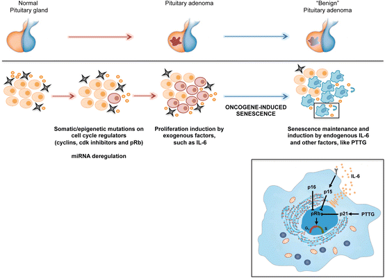
Pituitary adenomas are tumors that arise inside the pituitary gland. Pituitary adenomas are usually divided into 3 classes established upon their biological functioning: benign adenoma, invasive adenoma, and carcinomas. Most adenomas are benign, about 35% are invasive and simply 0.1% to 0.2% are carcinomas. Pituitary adenomas represent from 10% to 25% of all intracranial neoplasms and the anticipated incidence fee in the widespread populace is about 17%.

Non-invasive and non-secreting pituitary adenomas are taken into consideration to be benign in the literal as well as the medical experience; however a latest meta-evaluation (FernĂ¡ndez-Balsells, et al. 2011) of to be had studies has proven there are so far scant research – of poor satisfactory – to either help or refute this assumption.

Cause of Pinealoma

Cause of Pinealoma
Image Source-Google | Image by- | sciencedirect

Pinealomas can be due to proliferation of primary pineocytes (pineocytomas, pineoblastomas), astrocytes (astrocytoma), or germ cells (germinoma). Germinomas are the maximum commonplace tumor in the pineal gland.

Signs and symptoms of Pinealoma

Signs and symptoms of Pinealoma
Image Source-Google | Image by- | memorang

The pineal gland produces the hormone melatonin which performs a function in regulating circadian rhythms. A pinealoma may also disrupt production of this hormone, and insomnia can also result.

Frequently, paralysis of upward gaze at the side of several ocular findings inclusive of convergence retraction nystagmus and eyelid retraction additionally known as Collier's sign and Light Near Dissociation (student accommodates but doesn't react to light) are acknowledged collectively as Parinaud's syndrome or Dorsal Mid-brain syndrome, are the best bodily signs visible. This is caused by the compression of the vertical gaze center in the midbrain tectum at the level of the superior colliculus and cranial nerve III. Work-up usually consists of Neuro-imaging as seen at the proper.

A pinealoma may additionally motive interruption of hypothalamic inhibiting pathways, every so often leading to beta-hCG secretion and consequent Leydig's cell stimulation (endocrine syndrome).

Other signs and symptoms may include hydrocephalus, gait disturbances, and precocious puberty.

Pinealoma & it's Prognosis

Pinealoma & it's Prognosis
Image Source-Google | Image by- | healthhype
  • Pinealoma :

A pinealoma is a tumor of the pineal gland, part of the mind that produces melatonin. If a pinealoma destroys the cells of the pineal gland in a child, it can purpose precocious puberty.
  • Prognosis :

Of the exclusive types of pinealomas, the type with the most favorable analysis is the pineocytoma.

Prognosis of Oligodendroglioma

Prognosis of Oligodendroglioma
Image Source-Google | Image by- | cancer.gov

Oligodendrogliomas, like all different infiltrating gliomas, have a totally excessive (nearly uniform) charge of recurrence and step by step growth in grade over the years. Recurrent tumors are generally dealt with with more aggressive chemotherapy and radiation therapy. Recently, stereotactic surgical treatment has demonstrated a success in treating small tumors which have been identified early.

Long-time period survival is suggested in a minority of patients. With aggressive remedy and near tracking, it's far possible to survive the everyday lifestyles expectations for low grade oligodendroglioma. Westergaard's examine (1997) confirmed that patients younger than twenty years had a median survival of 17.5 years. For sufferers that are over 30, the survival fee is decrease however as remedy alternatives develop, the survival fee is higher. However, a affected person with bad fashionable health is more likely to pass sooner than those that have properly standard health.  Another study suggests a 34% survival rate after two decades. However, as mentioned above, such figures can be deceptive in view that they do now not issue in the styles of treatment nor the genetic signature of the tumors. Additionally, such historic information loses importance because of the notably long survival of patients (as compared to different kinds of mind tumors) and the introduction of more moderen treatment options through the years.

The standard dosing schedule of temozolomide

The standard dosing schedule of temozolomide
Image Source-Google | Image by- | researchgate

The trendy dosing schedule of temozolomide is five consecutive days of daily dosing at some stage in 28-day cycles. However, exceptional dosing schedules can also produce higher effects, together with non-stop every day dosing the use of decrease quantities of drug (e.G. 21-day dosing for the duration of 28-day cycles). As an instance of an altered dosing agenda, promising outcomes were proven the usage of decrease day by day doses on each day for 7 weeks, observed via a 4-week off periods. Regarding the duration of dosing, for oligodendrogliomas the duration prescribed by means of oncologists varies considerably and appears to range from 6 cycles to over 32 cycles (i.E. Over 3 years). In one have a look at, researchers in comparison sufferers who acquired temozolomide for as a minimum 12 months at the 5/28 day cycle, dividing such patients into two businesses: "quick time period" sufferers receiving temozolomide for 12–18 cycles and people "long term" sufferers receiving 19 or more cycles (range turned into 19 to 32 cycles). Researchers found that there was a statistically huge advantage for "long term" treatment (median development free survival for "quick term" patients changed into 95 weeks (comply with up of seventy three weeks), but for "long time" patients the median progression loose survival turned into now not yet reached (comply with up of 134 weeks)).

Because of their diffusely infiltrating nature, oligodendrogliomas cannot be absolutely resected and aren't curable via surgical excision. If the tumor mass compresses adjoining brain systems, a neurosurgeon will normally eliminate as a good deal of the tumor as he or she will be able to with out adverse other crucial, healthful mind systems. Surgery can be followed up via chemotherapy, radiation, or a mix of each, but current research advocate that radiation does not improve basic survival (even when age, medical records, histological grading, and form of surgical operation are considered).

Treatment of Oligodendroglioma


Treatment of Oligodendroglioma
Image Source-Google | Image by- | emedicinehealth


Oligodendrogliomas are normally felt to be incurable using present day remedies. However as compared to the extra not unusual astrocytomas, they're slowly growing with extended survival. In one series, median survival instances for oligodendrogliomas had been 11.6 years for grade II.
However, such figures can be misleading seeing that they do no longer issue inside the varieties of remedy nor the genetic signature of the tumors. A latest have a look at analyzed survival primarily based on chromosomal deletions and the consequences of radiation or chemotherapy as remedy, with the subsequent consequences (both low-grade and anaplastic oligodendrogliomas): 1p/19q deletion with radiation = 121 months (suggest), 1p/19q deletion with chemotherapy = over 160 months (imply now not yet reached), no 1p/19q deletion with radiation = 58 months (mean), and no 1p/19q deletion with chemotherapy = 75 months (suggest).
Because of the indolent nature of this tumor and the capability morbidity associated with neurosurgery, chemotherapy and radiation therapy, most neurooncologists will first of all pursue a direction of watchful waiting and deal with sufferers symptomatically. Symptomatic remedy frequently consists of the use of anticonvulsants for seizures and steroids for mind swelling.

Molecular genetics of Oligodendroglioma

Molecular genetics of Oligodendroglioma
Image Source-Google | Image by- | link.springer

By a ways, the most common structural deformity determined is co-deletion of chromosomal hands 1p and 19q. The excessive frequency of co-deletion is a hanging function of this glial tumor and is considered as a "genetic signature" of oligodendroglioma. Allelic losses on 1p and 19q, either one by one or combined, are greater commonplace in conventional oligodendrogliomas than in both astrocytomas or oligoastrocytomas. In one study, classic oligodendrogliomas showed 1p loss in 35 of 42 (83%) instances, 19q loss in 28 of 39 (72%), and these have been blended in 27 of 39 (69%) instances; there has been no extensive difference in 1p/19q loss of heterozygosity fame between low-grade and anaplastic oligodendroglioma. 1p/19q co-deletion has been correlated with each chemosensitivity and improved analysis in oligodendrogliomas. The gene products lost because of this codeletion may encompass mediators of resistance to genotoxic cures. Alternatively, 1p/19q loss might be an early oncogenic lesion selling the formation of glial neoplasms, which retain excessive sensitivity to genotoxic stress. Most large most cancers remedy centers mechanically take a look at for the deletion of 1p/19q as a part of the pathology document for oligodendrogliomas. The popularity of the 1p/19q loci may be detected through FISH, lack of heterozygosity (LOH) analysis or virtual karyotyping. Virtual karyotyping has the gain of assessing the complete genome in one assay, as well as the 1p/19q loci. This lets in assessment of other key loci in glial tumors, including EGFR and TP53 reproduction number reputation.

Whereas the prognostic relevance of 1p and 19q deletions is nicely mounted for mixed oligoastrocytomas, the prognostic relevance of the deletions for low-grade gliomas is more controversial. In terms of low-grade gliomas, a recent observe also suggests that 1p/19q co-deletion can be associated with a (1;19)(q10;p10) translocation which, like the blended 1p/19q deletion, is related to superior average survival and progression-unfastened survival in low-grade glioma patients. Oligodendrogliomas show simplest rarely mutations within the p53 gene, that is in evaluation to other gliomas. Epidermal increase component receptor amplification and whole 1p/19q codeletion are jointly unique and predictive of completely different outcomes, with EGFR amplification predicting poor analysis. There is a sturdy correlation between 1p/19q codeletion and the expression of proneural genes, suggesting that gliomas with a 1p19q codeletion constitute a subgroup of proneural gliomas.

Histopathological grading of Oligodendroglioma


Histopathological grading of Oligodendroglioma
Image Source-Google | Image by- | researchgate


The histopathologic grading of oligodendrogliomas is debatable. Currently, the most usually used grading schema is primarily based at the yr 2007 World Health Organization (WHO) suggestions. An up to date classification is in progress. Oligodendrogliomas are typically dichotomized into grade II (low grade) tumor.
Unfortunately, the WHO guidelines include subjective criteria in grade II. In addition, the presence of low mitotic interest, vascular proliferation and necrosis, along with pseudopallisading necrosis, is insufficient with the aid of itself to raise the grade of this tumor. This results in inevitable interobserver variability in prognosis by means of pathologists. The final responsibility for making remedy decisions and interpretation of this analysis lies with the oncologist in consultation with the patient and their family.
It has been proposed that WHO suggestions ought to incorporate a category for grade IV oligodendrogliomas which basically appear like glial neoplasms with overwhelming features of glioblastoma multiforme (GBM) bobbing up from acknowledged lower grade oligodendrogliomas or GBM with a sizeable proportion of oligodendroglial differentiation. The diagnostic software of this latter class is uncertain as these tumors may additionally behave both like glioblastoma or grade III Anaplastic oligodendrogliomas. As such,that is a really unusual prognosis.
The up to date WHO hints posted in 2007 recommends classifying such tumors at the moment as 'glioblastoma with oligodendroglioma component'. It stays to be mounted whether or no longer those tumors convey a higher diagnosis than trendy glioblastomas.

Microscopic appearance of Oligodendroglioma

Microscopic appearance of Oligodendroglioma
Image Source-Google | Image by- | emedicine.medscape


Oligodendrogliomas can't presently be differentiated from other brain lesions solely through their scientific or radiographic appearance. As such, a mind biopsy is the most effective approach of definitive prognosis. Oligodendrogliomas recapitulate the arrival of the ordinary resident oligodendroglia of the mind. (Their call derives from the Greek roots 'oligo' which means " few" and 'dendro' meaning "trees".) They are commonly composed of cells with small to slightly enlarged spherical nuclei with darkish, compact nuclei and a small amount of eosinophilic cytoplasm. They are regularly referred to as "fried egg" cells because of their histologic appearance. They appear as a humdrum population of mildly enlarged spherical cells infiltrating regular mind parenchyma and producing indistinct nodules. Although the tumor can also appear like vaguely circumscribed, it is by definition a diffusely infiltrating tumor.

Classically they generally tend to have a vasculature of finely branching capillaries that may take on a "hen twine" appearance. When invading gray count structures together with cortex, the neoplastic oligodendrocytes tend to cluster round neurons displaying a phenomenon called "perineuronal satellitosis". Oligodendrogliomas may also invade preferentially round vessels or under the pial floor of the brain.

Oligodendrogliomas have to be differentiated from the extra commonplace astrocytoma. Non-classical variations and combined tumors of each oligodendroglioma and astrocytoma differentiation are seen, making this distinction controversial among unique neuropathology agencies. In the United States, in wellknown, neuropathologists educated at the West Coast are extra liberal in the diagnosis of oligodendrogliomas than both East Coast or Midwest educated neuropathologists who render the diagnosis of oligodendroglioma for only traditional editions. Molecular diagnostics might also make this differentiation out of date within the destiny.

Other glial and glioneuronal tumors with which they may be frequently stressed due to their monotonous round cellular appearance consist of pilocytic astrocytoma, important neurocytoma, the so-referred to as dysembryoplastic neuroepithelial tumor, or occasionally ependymoma.

Cause of Oligodendroglioma

Cause of Oligodendroglioma
Image Source-Google | Image by- | in.pinterest

The purpose of oligodendrogliomas is unknown. Some studies have connected oligodendroglioma with a viral purpose. A 2009 Oxford Neurosymposium have a look at illustrated a 69% correlation among NJDS gene mutation and the tumor initiation. A single case file has related oligodendroglioma to irradiation of pituitary adenoma.

Signs and symptoms of Oligodendroglioma

Signs and symptoms of Oligodendroglioma
Image Source-Google | Image by- | researchgate

Oligodendroglioma get up specifically in the frontal lobe and in 50–80% of cases, the first symptom is the onset of seizure hobby, while not having any signs beforehand. Headaches blended with increased intracranial strain are also a not unusual symptom of oligodendroglioma. Depending on the region of the tumor many one-of-a-kind neurological deficits can be prompted, inclusive of, however not limited to, visual loss, motor weakness and cognitive decline. A computed tomography (CT) or magnetic resonance imaging (MRI) experiment is vital to represent tumor size, region, and hetero- or homogeneity. Final prognosis relies on biopsy and histopathologic examination of the tumor mass.

Oligodendroglioma

Oligodendroglioma
Image Source-Google | Image by- | sciencedirect

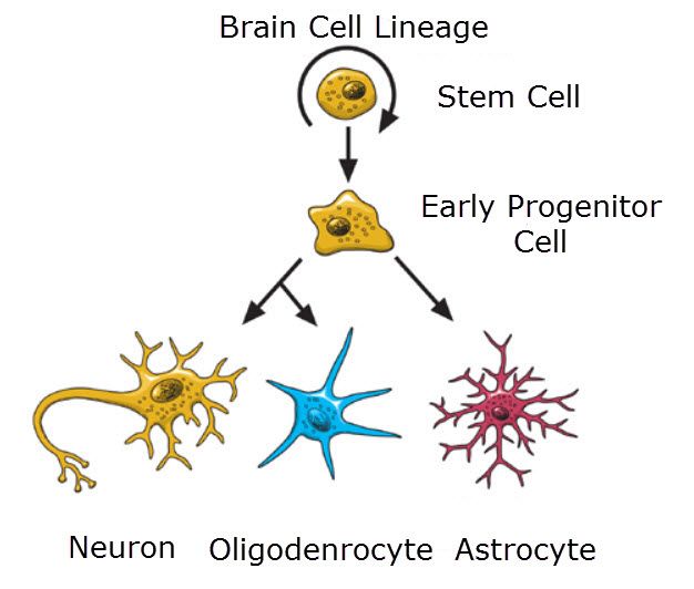
Oligodendrogliomas are a kind of glioma which might be believed to originate from the oligodendrocytes of the brain or from a glial precursor mobile. They occur commonly in adults (9.4% of all number one brain and principal anxious machine tumors) but also are observed in kids (4% of all primary mind tumors).

Refractory and relapsed neuroblastoma

Refractory and relapsed neuroblastoma
Image Source-Google | Image by- | broadcastmed

Chemotherapy with topotecan and cyclophosphamide is frequently used in refractory setting and after relapse.

A haploidentical stem cell transplant, that is, donor cells derived from parents, is being studied in those with refractory or relapsing neuroblastoma as stem cells from the person themselves is not useful.


Epidemiology of Neuroblastoma

Epidemiology of Neuroblastoma
Image Source-Google | Image by- | wikipedia

Neuroblastoma accommodates 6–10% of all formative years cancers, and 15% of cancer deaths in kids. The annual mortality rate is 10 in line with million youngsters within the 0- to 4-12 months-vintage age group, and four consistent with million inside the 4- to 9-yr vintage age group.

The maximum number of cases is within the 1st yr of lifestyles, and some cases are congenital. The age range is wide, consisting of older youngsters and adults, but most effective 10% of cases arise in humans older than 5 years of age. A massive European take a look at mentioned less than 2% of over 4000 neuroblastoma instances have been over 18 years antique.

Prognosis of Neuroblastoma

Prognosis of Neuroblastoma
Image Source-Google | Image by- | sciencedirect


By facts from England, the overall five-year survival price of neuroblastoma is 67%. Between 20% and 50% of high-threat cases do now not reply thoroughly to induction high-dose chemotherapy and are progressive or refractory. Relapse after of completion of frontline therapy is also not unusual. Further treatment is available in phase I and phase II clinical trials that test new dealers and combinations of retailers in opposition to neuroblastoma, but the final results stays very terrible for relapsed excessive-chance disease.

Most lengthy-term survivors alive today had low or intermediate threat sickness and milder guides of treatment as compared to high-danger disorder. The majority of survivors have lengthy-term results from the remedy. Survivors of intermediate and high-threat remedy often enjoy listening to loss, boom discount, thyroid function problems, getting to know difficulties, and greater chance of secondary cancers affect survivors of high-chance disease. An estimated 2 of 3 survivors of adolescence most cancers will in the long run expand at least one chronic and every so often life-threatening health hassle inside 20 to 30 years after the cancer diagnosis.


How does Chemotherapy used in the treatment of Neuroblastoma ?

How does Chemotherapy used in the treatment of Neuroblastoma ?
Image Source-Google | Image by- | intechopen

Chemotherapy dealers utilized in mixture had been discovered to be effective in opposition to neuroblastoma. Agents typically utilized in induction and for stem cellular transplant conditioning are platinum compounds (cisplatin, carboplatin), alkylating dealers (cyclophosphamide, ifosfamide, melphalan), topoisomerase II inhibitor (etoposide), anthracycline antibiotics (doxorubicin) and vinca alkaloids (vincristine). Some newer regimens consist of topoisomerase I inhibitors (topotecan and irinotecan) in induction that have been discovered to be effective in opposition to recurrent ailment.

In November 2020, naxitamab turned into accepted for scientific use in the United States in mixture with granulocyte-macrophage colony-stimulating element (GM-CSF) to treat people twelve months of age and older with excessive-chance neuroblastoma in bone or bone marrow whose tumor did no longer respond to or has come returned after previous treatments and has proven a partial reaction, minor response, or strong ailment to previous remedy.

People with low and intermediate risk disease of Neuroblastoma

People with low and intermediate risk disease of Neuroblastoma
Image Source-Google | Image by- | planetayurveda

People with low and intermediate hazard sickness have an terrific prognosis with treatment prices above 90% for low hazard and 70–90% for intermediate danger. In contrast, remedy for excessive-threat neuroblastoma the beyond  many years resulted in therapies only about 30% of the time. The addition of antibody therapy has raised survival fees for high-risk sickness extensively. In March 2009, an early evaluation of a Children's Oncology Group (COG) examine with 226 humans which might be high-hazard showed that  years after stem cell transplant 66% of the organization randomized to acquire ch14.18 antibody with GM-CSF and IL-2 were alive and sickness-unfastened compared to simplest 46% inside the organization that did no longer receive the antibody. The randomization became stopped so every body enrolling on the trial would acquire the antibody remedy.

Treatment of Neuroblastoma

Treatment of Neuroblastoma
Image Source-Google | Image by- | cancer

When the lesion is localized, it's far normally curable. However, long-time period survival for children with advanced disorder older than 18 months of age is bad despite aggressive multimodal remedy (intensive chemotherapy, surgical treatment, radiation therapy, stem mobile transplant, differentiation agent isotretinoin additionally called 13-cis-retinoic acid, and frequently immunotherapy with anti-GD2 monoclonal antibody therapy - dinutuximab).

Biologic and genetic characteristics have been recognized, which, while delivered to conventional scientific staging, has allowed undertaking to danger businesses for making plans remedy intensity. These criteria consist of the age of the person, volume of sickness unfold, microscopic look, and genetic functions along with DNA ploidy and N-myc oncogene amplification (N-myc regulates microRNAs), into low, intermediate, and high risk sickness. A latest biology look at (COG ANBL00B1) analyzed 2687 human beings with neuroblastoma and the spectrum of hazard assignment became decided: 37% of neuroblastoma instances are low chance, 18% are intermediate risk, and 45% are high risk. (There is some proof that the high- and occasional-danger sorts are because of one-of-a-kind mechanisms, and aren't merely two one of a kind levels of expression of the identical mechanism.)

The healing procedures for those distinctive threat classes are very unique.

Low-risk disease can frequently be discovered with none treatment in any respect or cured with surgical treatment on my own.

Intermediate-chance sickness is handled with surgical operation and chemotherapy.

High-risk neuroblastoma is treated with intensive chemotherapy, surgical treatment, radiation therapy, bone marrow / hematopoietic stem cellular transplantation, organic-based remedy with thirteen-cis-retinoic acid (isotretinoin or Accutane) and antibody remedy normally administered with the cytokines GM-CSF and IL-2. A meta analysis has determined proof that in children with excessive-danger neuroblastoma, remedy with myeloablative therapy improves event-free survival but can also boom the chance of side outcomes which includes kidney issues when in comparison to standard chemotherapy.

Diagnosis of Neuroblastoma

Diagnosis of Neuroblastoma
Image Source-Google | Image by- | labpedia

The analysis is usually showed by a surgical pathologist, taking into account the scientific presentation, microscopic findings, and other laboratory assessments. It can also get up from any neural crest element of the sympathetic frightened gadget (SNS).

Esthesioneuroblastoma, also known as olfactory neuroblastoma, is believed to rise up from the olfactory epithelium and its type stays controversial. However, since it is not a sympathetic nervous machine malignancy, esthesioneuroblastoma is a wonderful scientific entity and is not to be pressured with neuroblastoma.

Risk factors of Neuroblastoma

Risk factors of Neuroblastoma
Image Source-Google | Image by- | accbf

Several hazard factors were proposed and are the concern of ongoing research. Due to function early onset, many studies have centered on parental factors around conception and during gestation. Factors investigated have covered profession (i.E. Exposure to chemicals in precise industries), smoking, alcohol intake, use of medicinal tablets at some stage in pregnancy, and start factors; but, results have been inconclusive.

Other research have tested possible links with atopy and exposure to contamination early in existence, use of hormones and fertility drugs, and maternal use of hair dye.

Cause of Neuroblastoma

Cause of Neuroblastoma
Image Source-Google | Image by- | genengnews

The cause of neuroblastoma isn't nicely understood. The extremely good majority of cases are sporadic and nonfamilial. About 1–2% of cases run in households and were linked to specific gene mutations. Familial neuroblastoma in a few cases is due to uncommon germline mutations in the anaplastic lymphoma kinase (ALK) gene. Germline mutations within the PHOX2B or KIF1B gene were implicated in familial neuroblastoma, as well. Neuroblastoma is likewise a feature of neurofibromatosis type 1 and the Beckwith-Wiedemann syndrome.

MYCN oncogene amplification in the tumor is a common locating in neuroblastoma. The diploma of amplification indicates a bimodal distribution: either 3- to 10-fold, or a 100- to 300-fold. The presence of this mutation is fantastically correlated to superior degrees of disorder.

Duplicated segments of the LMO1 gene inside neuroblastoma tumor cells have been shown to increase the hazard of growing an aggressive shape of the most cancers.

Neuroblastoma has been linked to duplicate-wide variety variation in the NBPF10 gene, which results inside the 1q21.1 deletion syndrome or 1q21.1 duplication syndrome.

Most common location for neuroblastoma

Most common location for neuroblastoma
Image Source-Google | Image by- | mdpi

The maximum commonplace area for neuroblastoma to originate (i.E., the primary tumor) is inside the adrenal glands. This happens in 40% of localized tumors and in 60% of instances of big disease. Neuroblastoma can also broaden anywhere alongside the sympathetic frightened device chain from the neck to the pelvis. Frequencies in distinctive places consist of: neck (1%), chest (19%), abdomen (30% nonadrenal), or pelvis (1%). In rare cases, no number one tumor can be discerned.

Rare but feature displays encompass transverse myelopathy (tumor spinal twine compression, 5% of instances), treatment-resistant diarrhea (tumor vasoactive intestinal peptide secretion, 4% of cases), Horner's syndrome (cervical tumor, 2.4% of instances), opsoclonus myoclonus syndrome and ataxia (suspected paraneoplastic cause, 1.3% of cases), and hypertension (catecholamine secretion or kidney artery compression, 1.3% of instances).

Signs and symptoms of Neuroblastoma

Signs and symptoms of Neuroblastoma
Image Source-Google | Image by- | verywellhealth

The first symptoms of neuroblastoma are often indistinct, making prognosis tough. Fatigue, lack of appetite, fever, and joint pain are common. Symptoms rely on number one tumor locations and metastases if present:

  • In the abdomen, a tumor may also motive belly distension and constipation.
  • A tumor within the chest may additionally purpose respiration problems.
  • A tumor urgent at the spinal wire may reason weakness, hence an incapability to face, crawl, or stroll.
  • Bone lesions in the legs and hips may additionally purpose pain and limping.
  • A tumor in the bones around the eyes or orbits may purpose distinct bruising and swelling.
  • Infiltration of the bone marrow might also reason pallor from anemia.
Neuroblastoma regularly spreads to different parts of the frame before any signs are apparent, and 50 to 60% of all neuroblastoma cases gift with metastases.