Showing posts with label Deaths. Show all posts

Ependymoma tumors

Ependymoma tumors
Image Source-Google | Image by- | pinterest

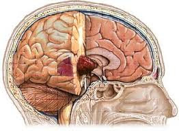
Ependymomas make up about 5% of person intracranial gliomas and up to ten% of adolescence tumors of the significant fearful system (CNS). Their prevalence seems to top at age five years and on the other hand at age 35. They increase from cells that line each the hollow cavities of the mind and the canal containing the spinal twine, but they generally rise up from the floor of the fourth ventricle, located inside the decrease back portion of the brain, in which they will produce headache, nausea and vomiting by obstructing the go with the flow of cerebrospinal fluid. This obstruction may purpose hydrocephalus. They may additionally rise up in the spinal wire, conus medullaris and supratentorial places. Other symptoms can include (but aren't restrained to): lack of appetite, issue slumbering, transient incapacity to distinguish colors, uncontrollable twitching, seeing vertical or horizontal strains whilst in vivid mild, and brief reminiscence loss. It need to be remembered that these signs are also popular in lots of different ailments now not associated with ependymoma.

Morphology of Ependymoma

Image Source-Google | Image by- | commons.wikimedia


Ependymomas are composed of cells with normal, spherical to oval nuclei. There is a variably dense fibrillary background. Tumor cells may additionally shape gland-like spherical or elongated systems that resemble the embryologic ependymal canal, with long, sensitive processes extending into the lumen; more regularly present are perivascular pseudorosettes wherein tumor cells are arranged around vessels with an intervening zone consisting of skinny ependymal strategies directed towards the wall of the vessel.

It has been suggested that ependymomas are derived from radial glia, despite their name suggesting an ependymal beginning.

Ependymoma

Ependymoma
Image Source-Google | Image by- | webpathology

An ependymoma is a tumor that arises from the ependyma, a tissue of the vital anxious machine. Usually, in pediatric cases the region is intracranial, even as in adults it's miles spinal. The not unusual area of intracranial ependymomas is the fourth ventricle. Rarely, ependymomas can arise inside the pelvic cavity.

Astrocytoma

Astrocytoma
Image Source-Google | Image by- | drprempillay

Astrocytomas are a kind of cerebrum cancer. They start in a specific sort of glial cells, star-formed synapses in the frontal cortex called astrocytes. This kind of growth doesn't generally spread external the mind and spinal string and it doesn't as a rule influence different organs. Astrocytomas are the most well-known glioma and can happen in many pieces of the cerebrum and infrequently in the spinal cord.

Hyperthermia of Malignancy (Cancer)

Hyperthermia of Malignancy (Treatment)
Image Source-Google | Image by- | verywellhealth


Malignancy can be treated using hyperthermia by applying either careful perfusion or interstitial procedures to the body. The utilization of this treatment type generally relies upon the way that dangerous and typical cells have varying reactions to the energy source used. This reliance is because of the intracellular changes which happen during hyperthermia; as the nucleic acids, cell film and cytoskeleton inside every phone is impacted by implication and additionally through different pathways. The blend of these intracellular changes implies there is no particular objective of cell demise in the hyperthermia process.

Post-Transplant Malignancy

Post-Transplant Malignancy
Image Source-Google | Image by- | ovariancancernewstoday

People who go through organ relocate a medical procedure have an expanded danger of creating threat in contrast with the general population. The most widely recognized type of harm being "nonmelanoma skin disease and, posttransplant lymphoproliferative disorders". The various sorts of harm created post-relocate rely upon which organ was transplanted. This is connected to beneficiaries being at a higher danger when presented to conventional danger factors just as, the sort and force of the activity, the span of their immunosuppression post-activity and, the danger of creating oncogenic viral infections.

Malignancy (CANCER)

Malignancy
Image Source-Google | Image by- | healio

Malignancy (from Latin male 'seriously', and - gnus 'conceived') is the propensity of an ailment to turn out to be logically more regrettable. 


Malignancy is generally recognizable as a portrayal of disease. A threatening growth diverges from a non-harmful harmless cancer in that a danger isn't self-restricted in its development, is fit for attacking into adjoining tissues, and might be equipped for spreading to far off tissues. A harmless growth has none of those properties.

NEOPLASM v/s TUMOR

NEOPLASM v/s TUMOR
Image Source-Google | Image by- | chapelboro


The word cancer or growth comes from the Latin word for expanding, which is one of the cardinal indications of aggravation. The word initially alluded to any type of expanding, neoplastic or not. In current English, cancer is utilized as an equivalent for neoplasm (a strong or liquid filled cystic injury that could possibly be framed by a strange development of neoplastic cells) that seems extended in size.Some neoplasms don't shape a growth - these remember leukemia and most types of carcinoma for situ. Growth is additionally not inseparable from disease. While disease is by definition dangerous, a cancer can be harmless, precancerous, or threatening.


The terms mass and knob are frequently utilized equivalently with cancer. As a rule, in any case, the term cancer is utilized conventionally, without reference to the actual size of the lesion. More explicitly, the term mass is frequently utilized when the sore has a maximal breadth of something like 20 millimeters (mm) in most noteworthy heading, while the term knob is typically utilized when the size of the injury is under 20 mm in its most noteworthy aspect (25.4 mm = 1 inch).

Abnormal growth of Tissues

Abnormal growth of Tissues
Image Source-Google | Image by- | science

Preceding the strange development of tissue, as neoplasia, cells regularly go through an unusual example of development, for example, metaplasia or dysplasia. However, metaplasia or dysplasia doesn't generally advance to neoplasia and can happen in different conditions as well. The word is from Ancient Greek νέος-neo 'new' and πλάσμα plasma 'arrangement, creation'.

NEOPLASM - CANCER

NEOPLASM - CANCER
Image Source-Google | Image by- | aacr


A neoplasm (/ˈniːoʊplæzəm, ˈniə-/) is a sort of unusual and inordinate development of tissue. The cycle that happens to frame or create a neoplasm is called neoplasia. The development of a neoplasm is awkward with that of the typical encompassing tissue, and continues developing strangely, regardless of whether the first trigger is removed. This unusual development ordinarily shapes a mass, when it very well might be known as a tumor.

Leiomyosarcoma (LMS) - Cancer

Image Source-Google | Image by- | bbc


Leiomyosarcoma is a dangerous (malignant) smooth muscle growth. A harmless growth beginning from a similar tissue is named leiomyoma. While leiomyosarcomas are not idea to emerge from leiomyomas, some leiomyoma variations' order is developing.

Treatment of Rhabdomyosarcoma (RMS)

Treatment of Rhabdomyosarcoma (RMS)
Image Source-Google | Image by- | moffitt


Therapy of Rhabdomyosarcoma (RMS), is a multidisciplinary work on including the utilization of medical procedure, chemotherapy, radiation, and perhaps immunotherapy. Medical procedure is for the most part the initial phase in a consolidated helpful methodology. Resectability changes relying upon growth site, and RMS frequently presents in locales that don't consider full careful resection without critical dismalness and loss of capacity. Under 20% of RMS growths are completely resected with negative edges. Rhabdomyosarcomas are profoundly chemosensitive, with roughly 80% of cases reacting to chemotherapy. Indeed, multi-specialist chemotherapy is demonstrated for all patients with rhabdomyosarcoma. Prior to the utilization of adjuvant and neoadjuvant treatment including chemotherapeutic specialists, treatment exclusively by careful means had an endurance pace of <20%. Present day endurance rates with adjuvant treatment are roughly 60–70%. 


There are two fundamental techniques for chemotherapy treatment for RMS. There is the VAC routine, comprising of vincristine, actinomycin D, and cyclophosphamide, and the IVA routine, comprising of ifosfamide, vincristine, and actinomycin D. These medications are managed in 9–15 cycles relying upon the organizing of the sickness and different treatments used. Other medication and treatment mixes may likewise show extra advantage. Expansion of doxorubicin and cisplatin to the VAC routine was displayed to build endurance paces of patients with alveolar-type, beginning phase RMS in IRS concentrate on III, and this equivalent expansion further developed endurance rates and multiplied bladder rescue rates in patients with stage III RMS of the bladder. In youngsters and youthful grown-ups with stage IV metastatic rhabdomyoscarcoma, a Cochrane audit has found no proof to help the utilization of high-portion chemotherapy as a standard therapy.


Radiation treatment, which kill malignant growth cells with centered portions of radiation, is regularly demonstrated in the therapy of rhabdomyosarcoma, and the prohibition of this therapy from infection the executives has been displayed to build repeat rates. Radiation treatment is utilized while resecting the whole of the cancer would include deformation or loss of significant organs (eye, bladder, and so forth) For the most part, regardless where an absence of complete resection is suspected, radiation treatment is indicated. Administration is normally following 6–12 weeks of chemotherapy if cancer cells are as yet present. The exemption for this timetable is the presence of parameningeal growths that have attacked the cerebrum, spinal string, or skull. In these cases radiation therapy is begun immediately. Now and again, uncommon radiation therapy might be required. Brachytherapy, or the position of little, radioactive "seeds" straightforwardly inside the growth or malignancy site, is regularly demonstrated in kids with cancers of delicate regions like the balls, bladder, or vagina. This decreases dispersing and the level of late poisonousness following dosing. Radiation treatment is all the more frequently showed in higher stage characterizations.

Signs and Symptoms of Rhabdomyosarcoma (RMS)

Signs and Symptoms of Rhabdomyosarcoma (RMS)
Image Source-Google | Image by- | cell


Rhabdomyosarcoma (RMS), can happen in practically any delicate tissue site in the body; the most well-known essential locales are genitourinary (24%), parameningeal (16%), furthest point (19%), circle (9%), other head and neck (10%), and incidental different destinations (22%). RMS regularly presents as a mass, however signs and indications can shift generally relying upon the site of the essential cancer. Genitourinary cancers might give hematuria, urinary parcel impediment, and additionally a scrotal or vaginal mass. Cancers that emerge in the retroperitoneum and mediastinum can turn out to be very enormous prior to creating signs and indications. Parameningeal cancers might give cranial nerve brokenness, manifestations of sinusitis, ear release, migraines, and facial agony. Orbital cancers frequently present with orbital enlarging and proptosis. Limit cancers by and large present as a quickly extending, firm mass in the applicable tissue. The malignancy's pervasiveness in the head, face, and neck will frequently take into consideration prior indications of the illness essentially because of the undeniable idea of growths in these locations. Despite the fluctuating show and normally forceful nature of the sickness, RMS can possibly be analyzed and treated early. The fourth IRSG investigation discovered that 23% of patients were analyzed on schedule for a total resection of their malignant growth, and 15% had resection with just insignificant remainders of the unhealthy cells.

TREATMENT OF OSTEOSARCOMA

A total extremist, careful, en coalition resection of the malignant growth, is the treatment of decision in osteosarcoma. Although around 90% of patients can have appendage rescue a medical procedure, difficulties, especially disease, prosthetic releasing and non-association, or neighborhood cancer repeat might cause the requirement for additional medical procedure or amputation.


TREATMENT OF OSTEOSARCOMA
Image Source-Google | Image by- | nature

Mifamurtide is utilized after a patient has had a medical procedure to eliminate the growth and along with chemotherapy to kill remaining malignancy cells to diminish the danger of disease repeat. Likewise, the choice to have rotation plasty after the growth is taken out exists.


Patients with osteosarcoma are best overseen by a clinical oncologist and a muscular oncologist experienced in overseeing sarcomas. Current standard treatment is to utilize neoadjuvant (chemotherapy given before a medical procedure) trailed by careful resection. The level of growth cell putrefaction (cell passing) found in the cancer after medical procedure gives a thought of the anticipation and furthermore informs the oncologist as to whether the chemotherapy routine ought to be adjusted after surgery.


Standard treatment is a blend of appendage rescue muscular medical procedure whenever the situation allows (or removal now and again) and a mix of high-portion methotrexate with leucovorin salvage, intra-blood vessel cisplatin, adriamycin, ifosfamide with mesna, BCD (bleomycin, cyclophosphamide, dactinomycin), etoposide, and muramyl tripeptide. Rotationplasty might be utilized. Ifosfamide can be utilized as an adjuvant treatment if the putrefaction rate is low.


Regardless of the accomplishment of chemotherapy for osteosarcoma, it has one of the least endurance rates for pediatric malignant growth. The best revealed 10-year endurance rate is 92%; the convention utilized is a forceful intra-blood vessel routine that individualizes treatment dependent on arteriographic response. Three-year occasion free endurance goes from half to 75%, and five-year endurance goes from 60% to 85+% in certain examinations. By and large, 65–70% patients treated five years prior will be alive today. These endurance rates are in general midpoints and shift significantly relying upon the singular corruption rate. 


Filgrastim or pegfilgrastim assist with white platelet counts and neutrophil counts. Blood bondings and epoetin alfa assist with sickliness. Computational examination on a board of osteosarcoma cell lines distinguished new shared and explicit restorative targets (proteomic and hereditary) in osteosarcoma, while aggregates showed an expanded job of growth microenvironments.

CAUSES OF OSTEOSARCOMA (OS) - CANCER

CAUSES OF OSTEOSARCOMA (CANCER)
Image Source-Google | Image by- | verywellhealth


A few exploration bunches are examining malignancy undifferentiated cells and their capability to cause growths alongside qualities and proteins causative in various phenotypes. Radiotherapy for random conditions might be an uncommon cause.

A little exaggerated marker chromosome or a goliath bar chromosome is available in the growth cells of poor quality OS including second rate focal OS and paraosteal OS (see beneath Variants section), convey different conceivably favorable to malignant qualities, and are thought to add to the improvement of these OS. Small effusive marker chromosomes and monster pole chromosomes in osteosarcomas.


CAUSES OF OSTEOSARCOMA (OS) - CANCER
Image Source-Google | Image by- | creativebiomart


Familial situations - where the cancellation of chromosome 13q14 inactivates the retinoblastoma quality is related with a high danger of osteosarcoma advancement. 

Bone dysplasias - including Paget's illness of bone, stringy dysplasia, enchondromatosis, and genetic various exostoses, increment the danger of osteosarcoma. 

Li–Fraumeni disorder (germline TP53 change) - is an inclining factor for osteosarcoma improvement. 

Rothmund – Thomson disorder (for example autosomal latent relationship of innate bone imperfections, hair and skin dysplasias, hypogonadism, and waterfalls) is related with expanded danger of this infection. 

Huge portions of Sr-90 - nicknamed bone searcher, expands the danger of bone disease and leukemia in creatures and is ventured to do as such in people.


There is no reasonable relationship between water fluoridation and malignant growth or passing because of disease, both for malignancy overall and furthermore explicitly for bone disease and osteosarcoma. Series of examination presumed that centralization of fluoride in water doesn't connect with osteosarcoma. The convictions in regards to relationship of fluoride openness and osteosarcoma come from an investigation of US National Toxicology program in 1990, which showed dubious proof of relationship of fluoride and osteosarcoma in male rodents. Be that as it may, there is still no strong proof of malignancy causing propensity of fluoride in mice. Fluoridation of water has been drilled all throughout the planet to work on residents' dental wellbeing. It is likewise considered as significant wellbeing success. Fluoride focus levels in water supplies are controlled, for example, United States Environmental Protection Agency directs fluoride levels to not be more noteworthy than 4 milligrams for each liter. Actually, water supplies as of now have normal happening fluoride, however numerous networks decided to add more fluoride to the point that it can diminish tooth decay. Fluoride is additionally known for its capacity to cause new bone formation. Yet, further examination shows no osteosarcoma hazards from fluoridated water in humans. Most of the exploration included counting number of osteosarcoma patients cases specifically regions which has contrast convergences of fluoride in drinking water. The measurement investigation of the information shows no huge distinction in events of osteosarcoma cases in various fluoridated regions. Another significant exploration included gathering bone examples from osteosarcoma patients to gauge fluoride fixation and contrast them with bone examples of recently analyzed threatening bone growths. The outcome is that the middle fluoride focuses in bone examples of osteosarcoma patients and cancer controls are not essentially different. Not just fluoride fixation in bones, Fluoride openings of osteosarcoma patients are likewise shown to be not fundamentally unique in relation to solid individuals. 

SIGNS AND SYMPTOMS OF OSTEOSARCOMA

Numerous patients initially whine of agony that might be more regrettable around evening time, might be discontinuous and of differing force and may have been happening for quite a while.


SIGNS AND SYMPTOMS OF OSTEOSARCOMA
Image Source-Google | Image by- | parenting.firstcry

Young people who are dynamic in sports frequently gripe of agony in the lower femur, or quickly underneath the knee. 


In the event that the growth is huge, it can present as unmistakable limited enlarging. Now and then an abrupt break is the primary indication on the grounds that the impacted bone isn't so solid as expected bone and may crack unusually with minor injury. In instances of all the more profound situated growths that are not as near the skin, like those starting in the pelvis, restricted enlarging may not be clear..

OSTEOSARCOMA - CANCER

OSTEOSARCOMA
Image Source-Google | Image by- | scopeblog


An osteosarcoma (OS) or osteogenic sarcoma (OGS) (or essentially bone malignancy), is a harmful growth in a bone. In particular, it is a forceful threatening neoplasm that emerges from crude changed cells of mesenchymal beginning (and along these lines a sarcoma) and that shows osteoblastic separation and produces harmful osteoid.

Osteosarcoma is the most widely recognized histological type of essential bone sarcoma. It is generally predominant in teens and youthful grown-ups.

MALIGNANT FIBROUS HISTIOCYTOMA - CANCER

MALIGNANT FIBROUS HISTIOCYTOMA
Image Source-Google | Image by- | google

MALIGNANT FIBROUS HISTIOCYTOMA, also known as UNDIFFERENTIATED PLEOMORPHIC SARCOMA, can be defined as soft-tissue sarcoma. 

Malignant Fibrous Histiocytoma is indicated a diagnoses of exclusion for sarcomas that cannot be more preciously classified or defined. Other sarcomas are cancer in the form of bone and soft-tissues, involving muscles, fat, blood-vessels and lymph vessels and fibrous tissues i.e., tendons and ligaments.

EWING'S SARCOMA - CANCER

EWING'S SARCOMA
Image Source-Google | Image by- | theconversation


EWING SARCOMA, can be defined as a kind of cancer that can be a bone sarcoma or a soft-tissue sarcoma. Signs and Symptoms may involve swelling and the pain at the site of tumor, fever and bone facture.

The most common areas where it begins are the legs, pelvis and the chest wall. Approximately, 25% of cases, in cancer has already spread and causes to other parts of the body at the times of diagnoses. Complications can involve a pleural effusion or paraplegia.

TYPES OF CANCER

TYPES OF CANCER
Image Source-Google | Image by- | fda.gov

  • BONE AND MUSCLE SARCOMA CANCER
  • BRAIN AND NERVOUS SYSTEM CANCER
  • BREAST CANCER
  • ENDOCRINE SYSTEM CANCER
  • EYE CANCER
  • GASTROINTESTINAL CANCER 
  • GENITOURINARY AND GYNECOLOGIC CANCER
  •  HEAD AND NECK CANCER
  • HEMATOPOIETIC CANCER
  • SKIN CANCER
  • THORACIC AND RESPIRATORY CANCER
  • HIV/AIDS CANCER