Cause of Pinealoma

Cause of Pinealoma
Image Source-Google | Image by- | sciencedirect

Pinealomas can be due to proliferation of primary pineocytes (pineocytomas, pineoblastomas), astrocytes (astrocytoma), or germ cells (germinoma). Germinomas are the maximum commonplace tumor in the pineal gland.

Signs and symptoms of Pinealoma

Signs and symptoms of Pinealoma
Image Source-Google | Image by- | memorang

The pineal gland produces the hormone melatonin which performs a function in regulating circadian rhythms. A pinealoma may also disrupt production of this hormone, and insomnia can also result.

Frequently, paralysis of upward gaze at the side of several ocular findings inclusive of convergence retraction nystagmus and eyelid retraction additionally known as Collier's sign and Light Near Dissociation (student accommodates but doesn't react to light) are acknowledged collectively as Parinaud's syndrome or Dorsal Mid-brain syndrome, are the best bodily signs visible. This is caused by the compression of the vertical gaze center in the midbrain tectum at the level of the superior colliculus and cranial nerve III. Work-up usually consists of Neuro-imaging as seen at the proper.

A pinealoma may additionally motive interruption of hypothalamic inhibiting pathways, every so often leading to beta-hCG secretion and consequent Leydig's cell stimulation (endocrine syndrome).

Other signs and symptoms may include hydrocephalus, gait disturbances, and precocious puberty.

Pinealoma & it's Prognosis

Pinealoma & it's Prognosis
Image Source-Google | Image by- | healthhype
  • Pinealoma :

A pinealoma is a tumor of the pineal gland, part of the mind that produces melatonin. If a pinealoma destroys the cells of the pineal gland in a child, it can purpose precocious puberty.
  • Prognosis :

Of the exclusive types of pinealomas, the type with the most favorable analysis is the pineocytoma.

Prognosis of Oligodendroglioma

Prognosis of Oligodendroglioma
Image Source-Google | Image by- | cancer.gov

Oligodendrogliomas, like all different infiltrating gliomas, have a totally excessive (nearly uniform) charge of recurrence and step by step growth in grade over the years. Recurrent tumors are generally dealt with with more aggressive chemotherapy and radiation therapy. Recently, stereotactic surgical treatment has demonstrated a success in treating small tumors which have been identified early.

Long-time period survival is suggested in a minority of patients. With aggressive remedy and near tracking, it's far possible to survive the everyday lifestyles expectations for low grade oligodendroglioma. Westergaard's examine (1997) confirmed that patients younger than twenty years had a median survival of 17.5 years. For sufferers that are over 30, the survival fee is decrease however as remedy alternatives develop, the survival fee is higher. However, a affected person with bad fashionable health is more likely to pass sooner than those that have properly standard health.  Another study suggests a 34% survival rate after two decades. However, as mentioned above, such figures can be deceptive in view that they do now not issue in the styles of treatment nor the genetic signature of the tumors. Additionally, such historic information loses importance because of the notably long survival of patients (as compared to different kinds of mind tumors) and the introduction of more moderen treatment options through the years.

The standard dosing schedule of temozolomide

The standard dosing schedule of temozolomide
Image Source-Google | Image by- | researchgate

The trendy dosing schedule of temozolomide is five consecutive days of daily dosing at some stage in 28-day cycles. However, exceptional dosing schedules can also produce higher effects, together with non-stop every day dosing the use of decrease quantities of drug (e.G. 21-day dosing for the duration of 28-day cycles). As an instance of an altered dosing agenda, promising outcomes were proven the usage of decrease day by day doses on each day for 7 weeks, observed via a 4-week off periods. Regarding the duration of dosing, for oligodendrogliomas the duration prescribed by means of oncologists varies considerably and appears to range from 6 cycles to over 32 cycles (i.E. Over 3 years). In one have a look at, researchers in comparison sufferers who acquired temozolomide for as a minimum 12 months at the 5/28 day cycle, dividing such patients into two businesses: "quick time period" sufferers receiving temozolomide for 12–18 cycles and people "long term" sufferers receiving 19 or more cycles (range turned into 19 to 32 cycles). Researchers found that there was a statistically huge advantage for "long term" treatment (median development free survival for "quick term" patients changed into 95 weeks (comply with up of seventy three weeks), but for "long time" patients the median progression loose survival turned into now not yet reached (comply with up of 134 weeks)).

Because of their diffusely infiltrating nature, oligodendrogliomas cannot be absolutely resected and aren't curable via surgical excision. If the tumor mass compresses adjoining brain systems, a neurosurgeon will normally eliminate as a good deal of the tumor as he or she will be able to with out adverse other crucial, healthful mind systems. Surgery can be followed up via chemotherapy, radiation, or a mix of each, but current research advocate that radiation does not improve basic survival (even when age, medical records, histological grading, and form of surgical operation are considered).

Treatment of Oligodendroglioma


Treatment of Oligodendroglioma
Image Source-Google | Image by- | emedicinehealth


Oligodendrogliomas are normally felt to be incurable using present day remedies. However as compared to the extra not unusual astrocytomas, they're slowly growing with extended survival. In one series, median survival instances for oligodendrogliomas had been 11.6 years for grade II.
However, such figures can be misleading seeing that they do no longer issue inside the varieties of remedy nor the genetic signature of the tumors. A latest have a look at analyzed survival primarily based on chromosomal deletions and the consequences of radiation or chemotherapy as remedy, with the subsequent consequences (both low-grade and anaplastic oligodendrogliomas): 1p/19q deletion with radiation = 121 months (suggest), 1p/19q deletion with chemotherapy = over 160 months (imply now not yet reached), no 1p/19q deletion with radiation = 58 months (mean), and no 1p/19q deletion with chemotherapy = 75 months (suggest).
Because of the indolent nature of this tumor and the capability morbidity associated with neurosurgery, chemotherapy and radiation therapy, most neurooncologists will first of all pursue a direction of watchful waiting and deal with sufferers symptomatically. Symptomatic remedy frequently consists of the use of anticonvulsants for seizures and steroids for mind swelling.

Molecular genetics of Oligodendroglioma

Molecular genetics of Oligodendroglioma
Image Source-Google | Image by- | link.springer

By a ways, the most common structural deformity determined is co-deletion of chromosomal hands 1p and 19q. The excessive frequency of co-deletion is a hanging function of this glial tumor and is considered as a "genetic signature" of oligodendroglioma. Allelic losses on 1p and 19q, either one by one or combined, are greater commonplace in conventional oligodendrogliomas than in both astrocytomas or oligoastrocytomas. In one study, classic oligodendrogliomas showed 1p loss in 35 of 42 (83%) instances, 19q loss in 28 of 39 (72%), and these have been blended in 27 of 39 (69%) instances; there has been no extensive difference in 1p/19q loss of heterozygosity fame between low-grade and anaplastic oligodendroglioma. 1p/19q co-deletion has been correlated with each chemosensitivity and improved analysis in oligodendrogliomas. The gene products lost because of this codeletion may encompass mediators of resistance to genotoxic cures. Alternatively, 1p/19q loss might be an early oncogenic lesion selling the formation of glial neoplasms, which retain excessive sensitivity to genotoxic stress. Most large most cancers remedy centers mechanically take a look at for the deletion of 1p/19q as a part of the pathology document for oligodendrogliomas. The popularity of the 1p/19q loci may be detected through FISH, lack of heterozygosity (LOH) analysis or virtual karyotyping. Virtual karyotyping has the gain of assessing the complete genome in one assay, as well as the 1p/19q loci. This lets in assessment of other key loci in glial tumors, including EGFR and TP53 reproduction number reputation.

Whereas the prognostic relevance of 1p and 19q deletions is nicely mounted for mixed oligoastrocytomas, the prognostic relevance of the deletions for low-grade gliomas is more controversial. In terms of low-grade gliomas, a recent observe also suggests that 1p/19q co-deletion can be associated with a (1;19)(q10;p10) translocation which, like the blended 1p/19q deletion, is related to superior average survival and progression-unfastened survival in low-grade glioma patients. Oligodendrogliomas show simplest rarely mutations within the p53 gene, that is in evaluation to other gliomas. Epidermal increase component receptor amplification and whole 1p/19q codeletion are jointly unique and predictive of completely different outcomes, with EGFR amplification predicting poor analysis. There is a sturdy correlation between 1p/19q codeletion and the expression of proneural genes, suggesting that gliomas with a 1p19q codeletion constitute a subgroup of proneural gliomas.

Histopathological grading of Oligodendroglioma


Histopathological grading of Oligodendroglioma
Image Source-Google | Image by- | researchgate


The histopathologic grading of oligodendrogliomas is debatable. Currently, the most usually used grading schema is primarily based at the yr 2007 World Health Organization (WHO) suggestions. An up to date classification is in progress. Oligodendrogliomas are typically dichotomized into grade II (low grade) tumor.
Unfortunately, the WHO guidelines include subjective criteria in grade II. In addition, the presence of low mitotic interest, vascular proliferation and necrosis, along with pseudopallisading necrosis, is insufficient with the aid of itself to raise the grade of this tumor. This results in inevitable interobserver variability in prognosis by means of pathologists. The final responsibility for making remedy decisions and interpretation of this analysis lies with the oncologist in consultation with the patient and their family.
It has been proposed that WHO suggestions ought to incorporate a category for grade IV oligodendrogliomas which basically appear like glial neoplasms with overwhelming features of glioblastoma multiforme (GBM) bobbing up from acknowledged lower grade oligodendrogliomas or GBM with a sizeable proportion of oligodendroglial differentiation. The diagnostic software of this latter class is uncertain as these tumors may additionally behave both like glioblastoma or grade III Anaplastic oligodendrogliomas. As such,that is a really unusual prognosis.
The up to date WHO hints posted in 2007 recommends classifying such tumors at the moment as 'glioblastoma with oligodendroglioma component'. It stays to be mounted whether or no longer those tumors convey a higher diagnosis than trendy glioblastomas.

Microscopic appearance of Oligodendroglioma

Microscopic appearance of Oligodendroglioma
Image Source-Google | Image by- | emedicine.medscape


Oligodendrogliomas can't presently be differentiated from other brain lesions solely through their scientific or radiographic appearance. As such, a mind biopsy is the most effective approach of definitive prognosis. Oligodendrogliomas recapitulate the arrival of the ordinary resident oligodendroglia of the mind. (Their call derives from the Greek roots 'oligo' which means " few" and 'dendro' meaning "trees".) They are commonly composed of cells with small to slightly enlarged spherical nuclei with darkish, compact nuclei and a small amount of eosinophilic cytoplasm. They are regularly referred to as "fried egg" cells because of their histologic appearance. They appear as a humdrum population of mildly enlarged spherical cells infiltrating regular mind parenchyma and producing indistinct nodules. Although the tumor can also appear like vaguely circumscribed, it is by definition a diffusely infiltrating tumor.

Classically they generally tend to have a vasculature of finely branching capillaries that may take on a "hen twine" appearance. When invading gray count structures together with cortex, the neoplastic oligodendrocytes tend to cluster round neurons displaying a phenomenon called "perineuronal satellitosis". Oligodendrogliomas may also invade preferentially round vessels or under the pial floor of the brain.

Oligodendrogliomas have to be differentiated from the extra commonplace astrocytoma. Non-classical variations and combined tumors of each oligodendroglioma and astrocytoma differentiation are seen, making this distinction controversial among unique neuropathology agencies. In the United States, in wellknown, neuropathologists educated at the West Coast are extra liberal in the diagnosis of oligodendrogliomas than both East Coast or Midwest educated neuropathologists who render the diagnosis of oligodendroglioma for only traditional editions. Molecular diagnostics might also make this differentiation out of date within the destiny.

Other glial and glioneuronal tumors with which they may be frequently stressed due to their monotonous round cellular appearance consist of pilocytic astrocytoma, important neurocytoma, the so-referred to as dysembryoplastic neuroepithelial tumor, or occasionally ependymoma.

Cause of Oligodendroglioma

Cause of Oligodendroglioma
Image Source-Google | Image by- | in.pinterest

The purpose of oligodendrogliomas is unknown. Some studies have connected oligodendroglioma with a viral purpose. A 2009 Oxford Neurosymposium have a look at illustrated a 69% correlation among NJDS gene mutation and the tumor initiation. A single case file has related oligodendroglioma to irradiation of pituitary adenoma.

Signs and symptoms of Oligodendroglioma

Signs and symptoms of Oligodendroglioma
Image Source-Google | Image by- | researchgate

Oligodendroglioma get up specifically in the frontal lobe and in 50–80% of cases, the first symptom is the onset of seizure hobby, while not having any signs beforehand. Headaches blended with increased intracranial strain are also a not unusual symptom of oligodendroglioma. Depending on the region of the tumor many one-of-a-kind neurological deficits can be prompted, inclusive of, however not limited to, visual loss, motor weakness and cognitive decline. A computed tomography (CT) or magnetic resonance imaging (MRI) experiment is vital to represent tumor size, region, and hetero- or homogeneity. Final prognosis relies on biopsy and histopathologic examination of the tumor mass.

Oligodendroglioma

Oligodendroglioma
Image Source-Google | Image by- | sciencedirect

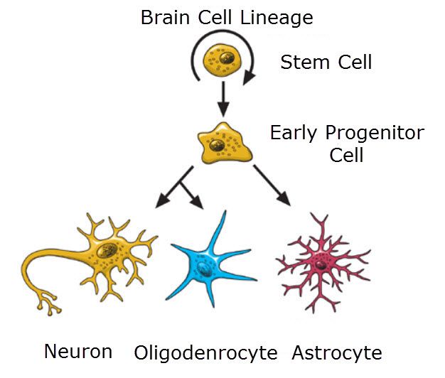
Oligodendrogliomas are a kind of glioma which might be believed to originate from the oligodendrocytes of the brain or from a glial precursor mobile. They occur commonly in adults (9.4% of all number one brain and principal anxious machine tumors) but also are observed in kids (4% of all primary mind tumors).

Refractory and relapsed neuroblastoma

Refractory and relapsed neuroblastoma
Image Source-Google | Image by- | broadcastmed

Chemotherapy with topotecan and cyclophosphamide is frequently used in refractory setting and after relapse.

A haploidentical stem cell transplant, that is, donor cells derived from parents, is being studied in those with refractory or relapsing neuroblastoma as stem cells from the person themselves is not useful.